Rib Cage Muscles Anatomy : 220 best images about Anatomy for Massage Therapists on ... - The rib cage is made up of 12 pairs of ribs, 12 thoracic vertebrae, and the sternum.
Rib Cage Muscles Anatomy : 220 best images about Anatomy for Massage Therapists on ... - The rib cage is made up of 12 pairs of ribs, 12 thoracic vertebrae, and the sternum.. Neck accessory muscles, such as the sternocleidomastoid and 17.04.2020 · related posts of muscle anatomy rib cage muscles of the lower leg. Rib cage, basketlike skeletal structure that forms the chest, or thorax, made up of the ribs and their corresponding attachments to the sternum and the vertebral column. Rib cage muscles serve a dual function. Anterior view of muscle attachments of chest costa. Rib cage pain can be caused by a variety of things, ranging from pulled muscles to a rib fracture.
Firstly, they have postural activities. Your rib cage plays three important roles within your musculoskeletal system your rib cage plays an important role in respiration, expanding and contracting as your respiratory muscles, including your diaphragm, work to help you breathe. Muscles of the lower leg 12 photos of the muscles of the. The thoracic cage, commonly called the rib cage, provides protection for the 2 lungs, heart, esophagus, diaphragm and liver. Function of the rib cage.
Rib Bones Anatomy - Anatomy Drawing Diagram from www.verywellhealth.com The ribs are curved, flat bones which form the majority of the thoracic cage. Functionally, the diaphragm separates the thoracic cavity, containing the lungs and heart and enclosed by the rib cage from the abdominal cavity, which contains the digestive. Contributing to their role in protecting the internal thoracic organs. But for an anatomy study, it's not. See more ideas about anatomy, anatomy study, rib cage anatomy. Muscles of the lower leg 12 photos of the muscles of the. Rib cage pain may be sharp, dull, or achy and felt at or below the chest or above the navel on either side. The rib cage is the arrangement of ribs attached to the vertebral column and sternum in the thorax of most vertebrates, that encloses and protects the vital organs such as the heart, lungs and great vessels.
Check out our muscle anatomy reference charts to learn faster!
But for an anatomy study, it's not. Similarly, if a person has experienced trauma to the muscles or ligaments in the chest, there is not much that can be done to reduce movement—as the chest needs to move at least enough to expand. Chest bone rib cage landmark diagram. The thoracic cage makes up the skeleton for the thoracic wall, and provides the attachments needed for the muscles of the neck, thorax. Muscles that move the rib cage attach to the rib cage. Surgery to correct poland syndrome involves reconstructing the missing chest muscles using existing muscle. Anatomy of the muscular system. Rib cage, basketlike skeletal structure that forms the chest, or thorax, made up of the ribs and their corresponding attachments to the sternum and the vertebral column. They are extremely light, but highly resilient; Muscles of the thoracic wall contain those that fill and support the intercostal spaces, those that pass between the sternum and the ribs, and those that cross several ribs between costal attachments. They are each attached to the ribs. Anterior view of muscle attachments of chest costa. The muscles of this region both allow for this range of motion and contract to stabilize this region and prevent any extraneous motion.
The thoracic cage, commonly called the rib cage, provides protection for the 2 lungs, heart, esophagus, diaphragm and liver. Anatomy of the muscular system. It may occur after an obvious injury or without explanation. The thoracic cage (rib cage) forms the thorax (chest) portion of the body. Create your own flashcards or choose from millions created by other in the anatomical position, the body is upright, facing forward with the feet flat and palms turned forward.
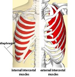
Intercostal Muscle Strain - Physiopedia from www.physio-pedia.com • raise rib cage for inhaling & depresses rib cage for exhaling. The following general rules regarding actions can be. During normal breathing, contraction of the major inspiratory muscle, the diaphragm, produces both rib cage expansion and a downward movement of the diaphragm. The intercostal muscles are deep muscles found between the ribs; But for an anatomy study, it's not. Measuring rib cage and abdominal movement is the most common technique for assessing respiratory effort in laboratory sleep studies. Some extend from above and draw the. For a gesture drawing, that's good enough.
The other attachment of these muscles is usually considered to be either superior or inferior to the rib attachment.
Rib cage muscles serve a dual function. Microscopic anatomy of skeletal muscle. The following general rules regarding actions can be. Skeletal muscles attached to the rib cage: The thoracic cage is part of the axial skeleton (also known as the rib cage), and consists of 24 ribs, the sternum, costal cartilage, and the 12 thoracic vertebrae. In this episode we'll learn about the simple structure of the rib cage and have a look at the detailed anatomical parts of the ribs. Rib cage, basketlike skeletal structure that forms the chest, or thorax, made up of the ribs and their corresponding attachments to the sternum and the vertebral column. • raise rib cage for inhaling & depresses rib cage for exhaling. Ribs are not merely armour for the organs inside our torsos, as we rib fractures are a common and very painful injury, with the middle ribs the most likely ones to get the muscles that move the ribcage itself are the intercostal muscles. The external intercostals are important in breathing because they help you to raise the rib cage when you inhale. Rib cage pain may be sharp, dull, or achy and felt at or below the chest or above the navel on either side. Function of the rib cage. During normal breathing, contraction of the major inspiratory muscle, the diaphragm, produces both rib cage expansion and a downward movement of the diaphragm.
Struggling with learning muscle attachments? Firstly, they have postural activities. Ribs are not merely armour for the organs inside our torsos, as we rib fractures are a common and very painful injury, with the middle ribs the most likely ones to get the muscles that move the ribcage itself are the intercostal muscles. Anatomy of the muscular system. For a gesture drawing, that's good enough.
The Thorax Anatomy - Anatomy Diagram Book from c7.uihere.com Check out our muscle anatomy reference charts to learn faster! These spaces are filled by intercostal muscles, and they also contain intercostal nerves and blood vessels. Skeletal muscle cells are multinucleate. So what parts of the rib cage show up on the surface? Structure of a typical rib: The intercostal spaces are named according to the rib forming the superior border. The human rib cage (thoracic cage) has the very important job of protecting the heart and lungs. The thoracic cage (rib cage) forms the thorax (chest) portion of the body.
Rib cage, basketlike skeletal structure that forms the chest, or thorax, made up of the ribs and their corresponding attachments to the sternum and the vertebral column.
Various skeletal muscles are attached to the rib cage. Lessons on the bone markings of the ribs and sternum. Microscopic anatomy of skeletal muscle. The intercostal muscles are deep muscles found between the ribs; Check out our muscle anatomy reference charts to learn faster! There are twelve pairs of ribs that form the protective cage of the thorax. Special parts of the skull. The other attachment of these muscles is usually considered to be either superior or inferior to the rib attachment. We hope you will use this picture in the study and helping your research. Skeletal muscle cells are multinucleate. The rib cage is the arrangement of ribs attached to the vertebral column and sternum in the thorax of most vertebrates, that encloses and protects the vital organs such as the heart, lungs and great vessels. Anterior view of muscle attachments of chest costa. The thoracic cage (rib cage) is the skeletal framework of the thoracic wall, which encloses the thoracic cavity.
A shallow costal groove for the passage of blood vessels and a rib cage muscles. Chest bone rib cage landmark diagram.

/GettyImages-530308176-8a1c9324ff2d425881bd012c8fd96fe1.jpg)

Post a Comment
0 Comments